Plasma Membrane and Transport - MCAT Biological and Biochemical Foundations of Living Systems
Card 1 of 392
Each of the following membrane transport processes requires the use of specific proteins that allow for movement across the plasma membrane EXCEPT .
Each of the following membrane transport processes requires the use of specific proteins that allow for movement across the plasma membrane EXCEPT .
Tap to reveal answer
Plasma membranes of the cell are permeable to molecules that pass through the phospholipid bilayer easily, namely small nonpolar molecules. Due to this specificity in permeability, membrane proteins are often required to transport molecules across the bilayer. Simple diffusion occurs when a substance passes through a membrane without the aid of an intermediary. All forms of facilitated transport, along with active transport, require the aid of specific membrane proteins. Thus, simple diffusion is the correct answer.
Plasma membranes of the cell are permeable to molecules that pass through the phospholipid bilayer easily, namely small nonpolar molecules. Due to this specificity in permeability, membrane proteins are often required to transport molecules across the bilayer. Simple diffusion occurs when a substance passes through a membrane without the aid of an intermediary. All forms of facilitated transport, along with active transport, require the aid of specific membrane proteins. Thus, simple diffusion is the correct answer.
← Didn't Know|Knew It →
Which of the following best describes the composition of the plasma membrane of an animal cell?
Which of the following best describes the composition of the plasma membrane of an animal cell?
Tap to reveal answer
The major components of the plasma membrane of an animal cell are lipids and proteins, with a small amount of carbohydrate components. The major lipid components are glycerophospholipids, sphingolipids, and some cholesterol. The amount of cholesterol varies depending upon certain factors, such as temperature, and helps maintain the fluidity of the membrane. Thus, the correct answer is phospholipids, sphingolipids, cholesterol, and protein, with some carbohydrate.
The major components of the plasma membrane of an animal cell are lipids and proteins, with a small amount of carbohydrate components. The major lipid components are glycerophospholipids, sphingolipids, and some cholesterol. The amount of cholesterol varies depending upon certain factors, such as temperature, and helps maintain the fluidity of the membrane. Thus, the correct answer is phospholipids, sphingolipids, cholesterol, and protein, with some carbohydrate.
← Didn't Know|Knew It →
What is the function of cholestrol in the cell plasma membrane?
What is the function of cholestrol in the cell plasma membrane?
Tap to reveal answer
The major purpose of cholestrol in the plasma membrane is to maintain membrane fluidity. Carbohydrates and glycoproteins function in cell-to-cell recognition, and proteins function in the transport of particles through the membrane. Charged particles can't freely pass through the membrane unless it is through a carrier protein.
The major purpose of cholestrol in the plasma membrane is to maintain membrane fluidity. Carbohydrates and glycoproteins function in cell-to-cell recognition, and proteins function in the transport of particles through the membrane. Charged particles can't freely pass through the membrane unless it is through a carrier protein.
← Didn't Know|Knew It →
Prions are the suspected cause of a wide variety of neurodegenerative diseases in mammals. According to prevailing theory, prions are infectious particles made only of protein and found in high concentrations in the brains of infected animals. All mammals produce normal prion protein, PrPC, a transmembrane protein whose function remains unclear.
Infectious prions, PrPRes, induce conformational changes in the existing PrPC proteins according to the following reaction:
PrPC + PrPRes → PrPRes + PrPRes
The PrPRes is then suspected to accumulate in the nervous tissue of infected patients and cause disease. This model of transmission generates replicated proteins, but does so bypassing the standard model of the central dogma of molecular biology. Transcription and translation apparently do not play a role in this replication process.
This theory is a major departure from previously established biological dogma. A scientist decides to test the protein-only theory of prion propagation. He establishes his experiment as follows:
Homogenized brain matter of infected rabbits is injected into the brains of healthy rabbits, as per the following table:
Rabbit 1 and 2: injected with normal saline on days 1 and 2
The above trials serve as controls.
Rabbit 3 and 4: injected with homogenized brain matter on days 1 and 2
The above trials use unmodified brain matter.
Rabbit 5 and 6: injected with irradiated homogenized brain matter on days 1 and 2
The above trials use brain matter that has been irradiated to destroy nucleic acids in the homogenate.
Rabbit 7 and 8: injected with protein-free centrifuged homogenized brain matter on days 1 and 2
The above trials use brain matter that has been centrifuged to generate a protein-free homogenate and a protein-rich homogenate based on molecular weight.
Rabbit 9 and 10: injected with boiled homogenized brain matter on days 1 and 2
The above trials use brain matter that have been boiled to destroy any bacterial contaminants in the homogenate.
Since PrPC is a transmembrane protein, what are we most likely to find in the part of the protein that spans the membrane?
Tap to reveal answer
The core of the lipid bilayer of all eukaryotic cells contains lipid; therefore, transmembrane proteins have a hydrophobic-rich series of residues in the area that spans the membrane.
The core of the lipid bilayer of all eukaryotic cells contains lipid; therefore, transmembrane proteins have a hydrophobic-rich series of residues in the area that spans the membrane.
← Didn't Know|Knew It →
Prions are the suspected cause of a wide variety of neurodegenerative diseases in mammals. According to prevailing theory, prions are infectious particles made only of protein and found in high concentrations in the brains of infected animals. All mammals produce normal prion protein, PrPC, a transmembrane protein whose function remains unclear.
Infectious prions, PrPRes, induce conformational changes in the existing PrPC proteins according to the following reaction:
PrPC + PrPRes → PrPRes + PrPRes
The PrPRes is then suspected to accumulate in the nervous tissue of infected patients and cause disease. This model of transmission generates replicated proteins, but does so bypassing the standard model of the central dogma of molecular biology. Transcription and translation apparently do not play a role in this replication process.
This theory is a major departure from previously established biological dogma. A scientist decides to test the protein-only theory of prion propagation. He establishes his experiment as follows:
Homogenized brain matter of infected rabbits is injected into the brains of healthy rabbits, as per the following table:
Rabbit 1 and 2: injected with normal saline on days 1 and 2
The above trials serve as controls.
Rabbit 3 and 4: injected with homogenized brain matter on days 1 and 2
The above trials use unmodified brain matter.
Rabbit 5 and 6: injected with irradiated homogenized brain matter on days 1 and 2
The above trials use brain matter that has been irradiated to destroy nucleic acids in the homogenate.
Rabbit 7 and 8: injected with protein-free centrifuged homogenized brain matter on days 1 and 2
The above trials use brain matter that has been centrifuged to generate a protein-free homogenate and a protein-rich homogenate based on molecular weight.
Rabbit 9 and 10: injected with boiled homogenized brain matter on days 1 and 2
The above trials use brain matter that have been boiled to destroy any bacterial contaminants in the homogenate.
A scientist realizes that the PrPC protein functions in normal cells to help regulate the cell membrane potential. Her research shows that cells with PrPC have a normal resting membrane potential at around –70 mV. Activation of PrPC causes depolarization, with a peak depolarization at around +60 mV. What ion, also present in action potentials, is PrPC most likely allowing to flow freely?
Tap to reveal answer
Students should know the main players in establishing action potentials are K+ and Na+. Further, Na+ inward flow through open channels brings an action potential to a peak depolarization of about +60 mV, which is sodium's equilibrium potential
Students should know the main players in establishing action potentials are K+ and Na+. Further, Na+ inward flow through open channels brings an action potential to a peak depolarization of about +60 mV, which is sodium's equilibrium potential
← Didn't Know|Knew It →
Prions are the suspected cause of a wide variety of neurodegenerative diseases in mammals. According to prevailing theory, prions are infectious particles made only of protein and found in high concentrations in the brains of infected animals. All mammals produce normal prion protein, PrPC, a transmembrane protein whose function remains unclear.
Infectious prions, PrPRes, induce conformational changes in the existing PrPC proteins according to the following reaction:
PrPC + PrPRes → PrPRes + PrPRes
The PrPRes is then suspected to accumulate in the nervous tissue of infected patients and cause disease. This model of transmission generates replicated proteins, but does so bypassing the standard model of the central dogma of molecular biology. Transcription and translation apparently do not play a role in this replication process.
This theory is a major departure from previously established biological dogma. A scientist decides to test the protein-only theory of prion propagation. He establishes his experiment as follows:
Homogenized brain matter of infected rabbits is injected into the brains of healthy rabbits, as per the following table:
Rabbit 1 and 2: injected with normal saline on days 1 and 2
The above trials serve as controls.
Rabbit 3 and 4: injected with homogenized brain matter on days 1 and 2
The above trials use unmodified brain matter.
Rabbit 5 and 6: injected with irradiated homogenized brain matter on days 1 and 2
The above trials use brain matter that has been irradiated to destroy nucleic acids in the homogenate.
Rabbit 7 and 8: injected with protein-free centrifuged homogenized brain matter on days 1 and 2
The above trials use brain matter that has been centrifuged to generate a protein-free homogenate and a protein-rich homogenate based on molecular weight.
Rabbit 9 and 10: injected with boiled homogenized brain matter on days 1 and 2
The above trials use brain matter that have been boiled to destroy any bacterial contaminants in the homogenate.
Another experiment shows that PrPC reacts with hormones that circulate among nervous tissue. As a transmembrane protein, what kinds of hormones are most likely to interact with PrPC?
I. Peptide hormones
II. Catecholamines
III. Steroid Hormones
Tap to reveal answer
Students should know that peptide hormones (and catecholamines, but this is not required to answer the question correctly as written here) interact with surface receptors and do not freely go through a membrane. They must interact with the transmembrane surface receptors to initiate a signal transduction cascade. In contrast, steroid hormones can bypass the transmembrance protein receptors by freely diffusing across the memberane, due to their small, nonpolar nature. In this case, only peptide hormones and catecholamines will require the facilitated diffusion mechanism provided by a transmembrane protein.
Students should know that peptide hormones (and catecholamines, but this is not required to answer the question correctly as written here) interact with surface receptors and do not freely go through a membrane. They must interact with the transmembrane surface receptors to initiate a signal transduction cascade. In contrast, steroid hormones can bypass the transmembrance protein receptors by freely diffusing across the memberane, due to their small, nonpolar nature. In this case, only peptide hormones and catecholamines will require the facilitated diffusion mechanism provided by a transmembrane protein.
← Didn't Know|Knew It →
The fluidity of plasma membranes .
The fluidity of plasma membranes .
Tap to reveal answer
Plasma membranes are composed of lipids and proteins, with a small amount of carbohydrates. The membrane is dependent upon these components to dictate its fluidity. An increase in unsaturated fatty acids leads to an increase in the fluidity of the membrane, while the increase of saturated fatty acids leads to a decrease in fluidity. Increasing the length of fatty acid chains leads to a decrease in fluidity. Thus, the correct answer is that it increases as the percent of unsaturated fatty acids increases.
Plasma membranes are composed of lipids and proteins, with a small amount of carbohydrates. The membrane is dependent upon these components to dictate its fluidity. An increase in unsaturated fatty acids leads to an increase in the fluidity of the membrane, while the increase of saturated fatty acids leads to a decrease in fluidity. Increasing the length of fatty acid chains leads to a decrease in fluidity. Thus, the correct answer is that it increases as the percent of unsaturated fatty acids increases.
← Didn't Know|Knew It →
The cell is the most basic functional unit of life. Everything that we consider to be living is made up of cells, and while there are different kinds of cells, they all have some essential features that link them all together under the category of "life." One of the most important parts of a cell is the membrane that surrounds it, seperating it from the rest of the environment.
While organisms from the three main domains live in incredibly different environments, they all possess similar cell membranes. This phospholipid bilayer protects the cell, giving it a way to allow certain things in while keeping other things out. Though organisms from different domains have different kinds of fatty linkages in their membranes, they all serve this essential purpose.
Membranes contain all kinds of essential proteins and signal molecules that allow the inside of the cell to respond to the outside of the cell. In a multicellular eukaryote, this ability can be used to allow cells to communicate. In a bacterial colony, an extracellular signal could be used to signal other bacteria. Signals cascade through a series of molecular pathways that go from the outside of the cell all the way to the nucleus and back out again, giving the cell control on a genetic level. This allows cellular responses to be quick and effective, and it also allows the cell to control how long it stays in that state.
Some proteins span the cellular membranes multiple times, weaving in and out of them. What parts of the protein would be on the inside and outside of the membrane?
Tap to reveal answer
The phospholipid bilayer is made of two layers. Each layer has hyrophilic heads facing outwards and hydrophobic tails facing inwards. So, the parts facing the inside and outside of the cell are hydrophilic and so hydrophilic parts of proteins would go there. The inside of the membrane is all long, saturated, fatty carbon tails that are hydrophilic would contain the hydrophilic portions of the protein. Like goes to like.
The phospholipid bilayer is made of two layers. Each layer has hyrophilic heads facing outwards and hydrophobic tails facing inwards. So, the parts facing the inside and outside of the cell are hydrophilic and so hydrophilic parts of proteins would go there. The inside of the membrane is all long, saturated, fatty carbon tails that are hydrophilic would contain the hydrophilic portions of the protein. Like goes to like.
← Didn't Know|Knew It →
The cell is the most basic functional unit of life. Everything that we consider to be living is made up of cells, and while there are different kinds of cells, they all have some essential features that link them all together under the category of "life." One of the most important parts of a cell is the membrane that surrounds it, seperating it from the rest of the environment.
While organisms from the three main domains live in incredibly different environments, they all possess similar cell membranes. This phospholipid bilayer protects the cell, giving it a way to allow certain things in while keeping other things out. Though organisms from different domains have different kinds of fatty linkages in their membranes, they all serve this essential purpose.
Membranes contain all kinds of essential proteins and signal molecules that allow the inside of the cell to respond to the outside of the cell. In a multicellular eukaryote, this ability can be used to allow cells to communicate. In a bacterial colony, an extracellular signal could be used to signal other bacteria. Signals cascade through a series of molecular pathways that go from the outside of the cell all the way to the nucleus and back out again, giving the cell control on a genetic level. This allows cellular responses to be quick and effective, and it also allows the cell to control how long it stays in that state.
One of the most important membrane proteins is the sodium-potassium pump. What would happen to a cell if this pump suddenly stopped working?
Tap to reveal answer
The sodium-potassium pump serves to move three sodium ions out of the cell and two potassium ion into the cell. These ions both have a plus one charge, so when the pump functions, it creates an environment where there are more solutes on the outside of the cell. if it stopped working, the cell would stop moving sodium out, and since it is a polar molecule, it can't cross the cell membrane on its own. There would be more solutes inside the cell than on the outside, and water would flow into the cell towards the higher solute concentration, causing the cell to swell and lyse.
The sodium-potassium pump serves to move three sodium ions out of the cell and two potassium ion into the cell. These ions both have a plus one charge, so when the pump functions, it creates an environment where there are more solutes on the outside of the cell. if it stopped working, the cell would stop moving sodium out, and since it is a polar molecule, it can't cross the cell membrane on its own. There would be more solutes inside the cell than on the outside, and water would flow into the cell towards the higher solute concentration, causing the cell to swell and lyse.
← Didn't Know|Knew It →
The sodium-potassium pump helps to maintain electrolyte gradients through use of ATP. Which of the following best describes this type of transport?
The sodium-potassium pump helps to maintain electrolyte gradients through use of ATP. Which of the following best describes this type of transport?
Tap to reveal answer
Active transport most correctly describes this type of movement, as it uses ATP as an energy source. In contrast, the other four choices are all different types of passive transport, constituting types of movement where no energy source is needed. Diffusion is simply the net movement of particles down their concentration gradient. Facilitated diffusion is a similar concept, but uses specialized transport proteins. Osmosis describes the movement of water, and lastly, filtration is the movement of both solute and water molecules.
Active transport most correctly describes this type of movement, as it uses ATP as an energy source. In contrast, the other four choices are all different types of passive transport, constituting types of movement where no energy source is needed. Diffusion is simply the net movement of particles down their concentration gradient. Facilitated diffusion is a similar concept, but uses specialized transport proteins. Osmosis describes the movement of water, and lastly, filtration is the movement of both solute and water molecules.
← Didn't Know|Knew It →
Scientists use a process called Flourescent In-Situ Hybridization, or FISH, to study genetic disorders in humans. FISH is a technique that uses spectrographic analysis to determine the presence or absence, as well as the relative abundance, of genetic material in human cells.
To use FISH, scientists apply fluorescently-labeled bits of DNA of a known color, called probes, to samples of test DNA. These probes anneal to the sample DNA, and scientists can read the colors that result using laboratory equipment. One common use of FISH is to determine the presence of extra DNA in conditions of aneuploidy, a state in which a human cell has an abnormal number of chromosomes. Chromosomes are collections of DNA, the totality of which makes up a cell’s genome. Another typical use is in the study of cancer cells, where scientists use FISH labels to ascertain if genes have moved inappropriately in a cell’s genome.
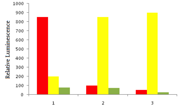
Using red fluorescent tags, scientists label probe DNA for a gene known to be expressed more heavily in cancer cells than normal cells. They then label a probe for an immediately adjacent DNA sequence with a green fluorescent tag. Both probes are then added to three dishes, shown below. In dish 1 human bladder cells are incubated with the probes, in dish 2 human epithelial cells are incubated, and in dish 3 known non-cancerous cells are used. The relative luminescence observed in regions of interest in all dishes is shown below.
The bladder cells in dish 1 begin to undergo programmed cell death, or apoptosis, when they initially become cancerous. If the cells form sodium-selective pores in their membranes to begin the process of cell death, sodium ions can begin to enter the cells without regulation. What will likely happen to a resting cell membrane potential when sodium enters?
Scientists use a process called Flourescent In-Situ Hybridization, or FISH, to study genetic disorders in humans. FISH is a technique that uses spectrographic analysis to determine the presence or absence, as well as the relative abundance, of genetic material in human cells.
To use FISH, scientists apply fluorescently-labeled bits of DNA of a known color, called probes, to samples of test DNA. These probes anneal to the sample DNA, and scientists can read the colors that result using laboratory equipment. One common use of FISH is to determine the presence of extra DNA in conditions of aneuploidy, a state in which a human cell has an abnormal number of chromosomes. Chromosomes are collections of DNA, the totality of which makes up a cell’s genome. Another typical use is in the study of cancer cells, where scientists use FISH labels to ascertain if genes have moved inappropriately in a cell’s genome.
Using red fluorescent tags, scientists label probe DNA for a gene known to be expressed more heavily in cancer cells than normal cells. They then label a probe for an immediately adjacent DNA sequence with a green fluorescent tag. Both probes are then added to three dishes, shown below. In dish 1 human bladder cells are incubated with the probes, in dish 2 human epithelial cells are incubated, and in dish 3 known non-cancerous cells are used. The relative luminescence observed in regions of interest in all dishes is shown below.
The bladder cells in dish 1 begin to undergo programmed cell death, or apoptosis, when they initially become cancerous. If the cells form sodium-selective pores in their membranes to begin the process of cell death, sodium ions can begin to enter the cells without regulation. What will likely happen to a resting cell membrane potential when sodium enters?
Tap to reveal answer
The pores formed are, according to the question, sodium selective. So it is unlikely that potassium concentration changes will be a major contributor to membrane potential changes. Since sodium is postively charged, and the ions entering are sodium, the inside of the cell will become more positively charged as sodium permeability goes up. We know that sodium will enter and potassium will leave due to the established gradients determined by sodium-potassium ATPase.
The pores formed are, according to the question, sodium selective. So it is unlikely that potassium concentration changes will be a major contributor to membrane potential changes. Since sodium is postively charged, and the ions entering are sodium, the inside of the cell will become more positively charged as sodium permeability goes up. We know that sodium will enter and potassium will leave due to the established gradients determined by sodium-potassium ATPase.
← Didn't Know|Knew It →
Cryptosporidium is a genus of gastrointestinal parasite that infects the intestinal epithelium of mammals. Cryptosporidium is water-borne, and is an apicomplexan parasite. This phylum also includes Plasmodium, Babesia, and Toxoplasma.
Apicomplexans are unique due to their apicoplast, an apical organelle that helps penetrate mammalian epithelium. In the case of cryptosporidium, there is an interaction between the surface proteins of mammalian epithelial tissue and those of the apical portion of the cryptosporidium infective stage, or oocyst. A scientist is conducting an experiment to test the hypothesis that the oocyst secretes a peptide compound that neutralizes intestinal defense cells. These defense cells are resident in the intestinal epithelium, and defend the tissue by phagocytizing the oocysts.
She sets up the following experiment:
As the neutralizing compound was believed to be secreted by the oocyst, the scientist collected oocysts onto growth media. The oocysts were grown among intestinal epithelial cells, and then the media was collected. The media was then added to another plate where Toxoplasma gondii was growing with intestinal epithelial cells. A second plate of Toxoplasma gondii was grown with the same type of intestinal epithelium, but no oocyst-sourced media was added.
The apicoplast that defines the phylum Apicomplexa is a membrane bound organelle. Which of the following is true of membrane-bound organelles?
I. They are only present in eukaryotes
II. They are bound by a single phospholipid layer
III. They do not have membrane-associated proteins attached
Tap to reveal answer
Membrane-bound organelles are a key distinction between eukaryotic cells and prokaryotic cells. Membrane-bound organelles serve diverse purposes, and often have associated protein structures to help carry out enzymatic reactions or other functions.
Cell membranes are almost invariably at least bilayers, however, making choice 2 incorrect. A bilayer functions to sequester the lipid tails common to membranes away from the aqueous cytosol. Incidentally, the apicoplast is surrounded by four membranes, but the effect is the same.
← Didn't Know|Knew It →
Cryptosporidium is a genus of gastrointestinal parasite that infects the intestinal epithelium of mammals. Cryptosporidium is water-borne, and is an apicomplexan parasite. This phylum also includes Plasmodium, Babesia, and Toxoplasma.
Apicomplexans are unique due to their apicoplast, an apical organelle that helps penetrate mammalian epithelium. In the case of cryptosporidium, there is an interaction between the surface proteins of mammalian epithelial tissue and those of the apical portion of the cryptosporidium infective stage, or oocyst. A scientist is conducting an experiment to test the hypothesis that the oocyst secretes a peptide compound that neutralizes intestinal defense cells. These defense cells are resident in the intestinal epithelium, and defend the tissue by phagocytizing the oocysts.
She sets up the following experiment:
As the neutralizing compound was believed to be secreted by the oocyst, the scientist collected oocysts onto growth media. The oocysts were grown among intestinal epithelial cells, and then the media was collected. The media was then added to another plate where Toxoplasma gondii was growing with intestinal epithelial cells. A second plate of Toxoplasma gondii was grown with the same type of intestinal epithelium, but no oocyst-sourced media was added.
Upon studying cryptosporidium under a microscope, the scientist in the passages notices that each oocyst has a thick cell wall. When the scientist puts the oocysts into pure water, the oocysts remain intact while other cells swell. Against what kind of stress is the oocyst cell wall protecting?
Tap to reveal answer
As described in the question, the shell is protecting against osmotic stress. We would expect osmotic stress to be present when we put the oocysts in pure water, which is hypotonic relative to the oocyst itself. The tendency of non-protected cells to burst in such a condition is the result of osmotic stress caused by the influx of water diffusing across the membrane.
As described in the question, the shell is protecting against osmotic stress. We would expect osmotic stress to be present when we put the oocysts in pure water, which is hypotonic relative to the oocyst itself. The tendency of non-protected cells to burst in such a condition is the result of osmotic stress caused by the influx of water diffusing across the membrane.
← Didn't Know|Knew It →
What is the average resting potential of a nerve cell membrane?
What is the average resting potential of a nerve cell membrane?
Tap to reveal answer
Membrane potential is the difference between the electric potential inside the cell and the electric potential outside the cell. At rest, the membrane potential of most cells (including nerve cells) is between -70mV and -80mV due to the concentration of intracellular and extracellular potassium and sodium ions. The expulsion of sodium ions, in particular, contributes to positive charges outside the cell and lowers the charge inside.
Membrane potential is the difference between the electric potential inside the cell and the electric potential outside the cell. At rest, the membrane potential of most cells (including nerve cells) is between -70mV and -80mV due to the concentration of intracellular and extracellular potassium and sodium ions. The expulsion of sodium ions, in particular, contributes to positive charges outside the cell and lowers the charge inside.
← Didn't Know|Knew It →
Which of the following are true about a cell's phospholipid bilayer?
Which of the following are true about a cell's phospholipid bilayer?
Tap to reveal answer
Each phospholipid consists of a polar phosphate head and two non-polar lipid tails. The phospholipid bilayer consists of polar heads facing the inside and outside of the cell, which interact with polar, aqueous environments. The non-polar hydrocarbon tails are packed on the inside of the bilayer, as far away from the polar environments as possible.
Each phospholipid consists of a polar phosphate head and two non-polar lipid tails. The phospholipid bilayer consists of polar heads facing the inside and outside of the cell, which interact with polar, aqueous environments. The non-polar hydrocarbon tails are packed on the inside of the bilayer, as far away from the polar environments as possible.
← Didn't Know|Knew It →
Each answer choice below contains two modes of cellular transport. Select the choice in which both modes are passive.
Each answer choice below contains two modes of cellular transport. Select the choice in which both modes are passive.
Tap to reveal answer
Facilitated diffusion and osmosis are both forms of passive transport. Broadly, diffusion is defined as the movement of any substance from a higher to a lower concentration along a gradient. This definition includes osmosis, which is the diffusion of water.
Endocytosis, exocytosis, and pinocytosis are all modes of bulk transport and require energy. Similarly, the sodium-potassium and proton pumps are forms of active transport.
← Didn't Know|Knew It →
Cells can acquire substances from the extracellular environment through a process called endocytosis, of which there are multiple subtypes.
Which of the following describes the process by which extracellular fluid is taken up by small invaginations of the cell membrane?
Cells can acquire substances from the extracellular environment through a process called endocytosis, of which there are multiple subtypes.
Which of the following describes the process by which extracellular fluid is taken up by small invaginations of the cell membrane?
Tap to reveal answer
Pinocytosis, or "cell drinking", refers to the process by which extracellular fluids are engulfed by invaginations of the cellular membrane to create vesicles of the fluid. Phagocytosis is a similar mechanism that acquires extracellular solids into the cell. Receptor-mediated endocytosis occurs when hormones and nutrients bind to a ligand-specific receptor on the cellular membrane and clathrin-mediated endocytosis involves entrance to the cell via clathrin-coated pits.
Pinocytosis, or "cell drinking", refers to the process by which extracellular fluids are engulfed by invaginations of the cellular membrane to create vesicles of the fluid. Phagocytosis is a similar mechanism that acquires extracellular solids into the cell. Receptor-mediated endocytosis occurs when hormones and nutrients bind to a ligand-specific receptor on the cellular membrane and clathrin-mediated endocytosis involves entrance to the cell via clathrin-coated pits.
← Didn't Know|Knew It →
One component of the immune system is the neutrophil, a professional phagocyte that consumes invading cells. The neutrophil is ferried to the site of infection via the blood as pre-neutrophils, or monocytes, ready to differentiate as needed to defend their host.
In order to leave the blood and migrate to the tissues, where infection is active, the monocyte undergoes a process called diapedesis. Diapedesis is a process of extravasation, where the monocyte leaves the circulation by moving in between endothelial cells, enters the tissue, and matures into a neutrophil.
Diapedesis is mediated by a class of proteins called selectins, present on the monocyte membrane and the endothelium. These selectins interact, attract the monocyte to the endothelium, and allow the monocytes to roll along the endothelium until they are able to complete diapedesis by leaving the vasculature and entering the tissues.
The image below shows monocytes moving in the blood vessel, "rolling" along the vessel wall, and eventually leaving the vessel to migrate to the site of infection.

The movement of monocytes between endothelial cells can best be characterized as .
One component of the immune system is the neutrophil, a professional phagocyte that consumes invading cells. The neutrophil is ferried to the site of infection via the blood as pre-neutrophils, or monocytes, ready to differentiate as needed to defend their host.
In order to leave the blood and migrate to the tissues, where infection is active, the monocyte undergoes a process called diapedesis. Diapedesis is a process of extravasation, where the monocyte leaves the circulation by moving in between endothelial cells, enters the tissue, and matures into a neutrophil.
Diapedesis is mediated by a class of proteins called selectins, present on the monocyte membrane and the endothelium. These selectins interact, attract the monocyte to the endothelium, and allow the monocytes to roll along the endothelium until they are able to complete diapedesis by leaving the vasculature and entering the tissues.
The image below shows monocytes moving in the blood vessel, "rolling" along the vessel wall, and eventually leaving the vessel to migrate to the site of infection.
The movement of monocytes between endothelial cells can best be characterized as .
Tap to reveal answer
Paracellular transport moves material between cells, while transcellular transport moves things through cells; thus, this is an example of paracellular transport.
Facilitated diffusion, pinocytosis, and passive transport all involve the entrance of a substance into a cell. Monocytes are transferring location, but are not entering another cell in the process.
← Didn't Know|Knew It →
When a solute moves down its concentration gradient across a non-permeable barrier, the process is known as .
When a solute moves down its concentration gradient across a non-permeable barrier, the process is known as .
Tap to reveal answer
A solute moving down its concentration gradient across a non-permeable barrier is an example of facilitated diffusion. It requires a carrier protein, but no energy. Any particle crossing a non-permeable barrier will require a protein, and cannot pass via diffusion or osmosis. ATP will not be required to transport a particle down its gradient.
If the particle were travelling against its gradient, it would require ATP AND a protein, and active transport would be the correct answer. Simple diffusion and osmosis require no energy or protein.
← Didn't Know|Knew It →
Which of the following is generally permeable to the cell membrane?
Which of the following is generally permeable to the cell membrane?
Tap to reveal answer
Albumin, glucose, and potassium ions are all examples of NON-permeable solutes. To be permeable, a solute must be small and nonpolar. Albumin, the main osmoregulatory protein, is bulky and too large to cross the membrane freely. Glucose is also large, as well as polar, and cannot cross the membrane. Potassium cannot freely cross due to the positive charge on the ion. All of these will require facilitated means of entering the cell.
Testosterone is a steroid hormone, and as such is small and nonpolar. All steroid hormones have intracellular receptors, and are able to enter a cell freely. Of the four choices, it is the only solute that can permeate the cell membrane.
← Didn't Know|Knew It →